
Antibodies
| Rabbit polyclonal antibody to SMAD-2 | |
|---|---|
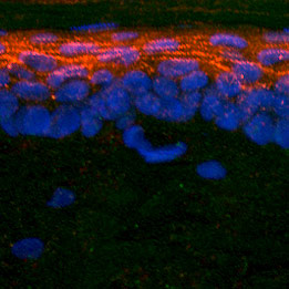 Immunohistochemical detection of SMAD-2 in mouse skin. Tissue was fixed with 4% formaldehyde and cut into 10 μm thick cryostat sections. Tissue was incubated with rabbit polyclonal antibody to SMAD-2 at 12 μg/mL overnight at 4°C followed by incubation with Donkey anti-rabbit Rhodamine Red conjugated secondary antibodies at 1:200. Immunohistochemical detection of SMAD-2 in mouse skin. Tissue was fixed with 4% formaldehyde and cut into 10 μm thick cryostat sections. Tissue was incubated with rabbit polyclonal antibody to SMAD-2 at 12 μg/mL overnight at 4°C followed by incubation with Donkey anti-rabbit Rhodamine Red conjugated secondary antibodies at 1:200. Nuclei of keratinocytes were counterstained with DAPI (blue) |
 |
| Formulation | Lyophilized powder |
| Purification | Affinity purified |
| Host Species | Rabbit |
| Unit Size: | 50 µg |
| Immunogen | Synthetic peptide |
| Sequence: | MSSILPFTPPVVKRLLGWKKSAGGSGGAGGGEQNGQEEKWCEKAVKSLVK |
| Alternative Names | JV18-1, Mad-related protein 2. |
| Accession Number: | Q15796 |
| Gene Symbol | SMAD2 |
| Accession URL: | http://www.uniprot.org/uniprot/Q15796 |
| Function: SMAD2 belongs to the SMAD, a family of proteins similar to the gene products of the Drosophila gene 'mothers against decapentaplegic' (Mad) and the C. elegans gene Sma. SMAD proteins are signal transducers and transcriptional modulators that mediate multiple signaling pathways. SMAD2 mediates effects of the transforming growth factor (TGF)-beta, and thus regulates multiple cellular processes, such as cell proliferation, apoptosis, and differentiation. SMAD2 is recruited to the TGF-beta receptors through its interaction with the SMAD anchor for receptor activation (SARA) protein. In response to TGF-beta signal, SMAD2 is phosphorylated by the TGF-beta receptors. The phosphorylation induces the dissociation of this protein with SARA and the association with the family member SMAD4. The association with SMAD4 is important for the translocation of this protein into the nucleus, where it binds to target promoters and forms a transcription repressor complex with other cofactors. |
|
| Applications: | Immunohistochemistry (IHC), Immunocytochemistry (ICC), Western Blotting (WB). |
| Working Dilution for Immunofluorescence (ICC): | 5 – 15 µg/mL |
| Working Dilution for Immunohistochemistry (IHC): | 5 – 10 µg/mL |
| Working Dilution for Western Blottin (WB): | 1 µg/mL |
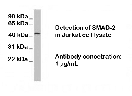
| IHC Positive control: | Skin, Jurkat cell line. |
| Specificity: | Confirmed by WB. |
| Cross-reactivity: | Human; mouse; rat |
| Reconstitution: | Reconstitute in 0.05 mL of PBS (pH 7.4) to achieve an antibody concentration of 1000 µg/mL. Centrifuge to remove any insoluble material. |
| Storage / Stability: | At least 12 months after purchase at 2 - 4°C. After reconstitution, aliquot and store at -20°C for a higher stability and at 4°C with an appropriate antibacterial agent. Avoid freeze-thaw cycles. |
References
|
|